
Granty wyjazdowe na Kongres ICE Thessaloniki 2025!
Szanowni Państwo,
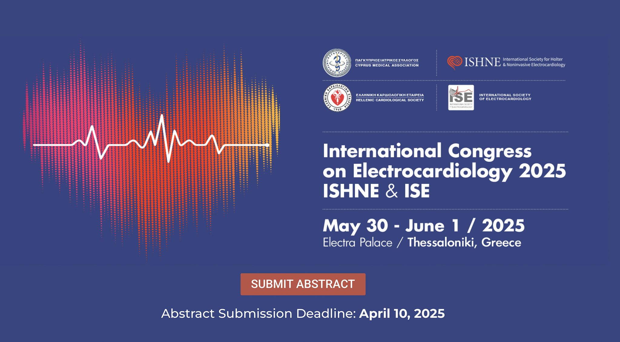
W dniach 30 maja - 01 czerwca 2025 w greckich Salonikach odbędzie się kongres ICE (International Congress on Electrocardiology), organizowany wspólnie przez dwa towarzystwa naukowe: International Society of Electrocardiology (ISE) oraz International Society for Holter and Noninvasive Electrocardiology (ISHNE).
Zarząd AENiT podjął decyzję o ufundowaniu grantów wyjazdowych w kwocie do 3000 PLN dla aktywnych uczestników tego kongresu (wykładowców, osób prowadzących sesje lub pierwszych autorów prac oryginalnych, które zostały przyjęte do prezentacji).
Zapraszamy do przysyłania zgłoszeń do 18 maja 2025 na adres mailowy: sekretarz_aenit@ptkardio.pl
Aplikacja musi zawierać dane osobowe oraz dowód wykładu/prezentacji/prowadzenia sesji w czasie kongresu ICE 2025.
Osoby ubiegające się o grant wyjazdowy muszą być członkami AENiT i mieć opłacone składki członkowskie PTK.
Link do strony kongresu: https://ishne2025.frei.gr/
Z wyrazami szacunku,
Olgierd Woźniak,
Sekretarz AENiT PTK
Aktualności

Zapraszamy do zapoznania się z artykułami opracowanymi w ramach Biuletynu AENiT
W cyklicznych Biuletynach AENiT będziemy starali się przedstawiać najważniejsze informacje z życia Asocjacji.

Granty wyjazdowe dla aktywnych uczestników kongresu ISHNE w Tesalonikach
Granty wyjazdowe dla aktywnych uczestników kongresu ISHNE w Tesalonikach
Zarząd AENiT w kadencji 2023-2025
Przewodnicząca
prof. dr hab. n. med.
Ewa Piotrowicz
Kierownik Centrum Telekardiologii
Narodowy Instytut Kardiologii
Stefana Kardynała Wyszyńskiego,
Państwowy Instytut Badawczy
Przewodnicząca-Elekt
prof. dr hab. n. med.
Małgorzata Kurpesa
Katedra i I Klinika Kardiologii Uniwersytetu
Medycznego w Łodzi,
Oddział Rehabilitacji Kardiologicznej
WSSZ. im. W. Biegańskiego w Łodzi
Poprzednia Przewodnicząca
prof. dr hab. n. med.
Elżbieta Katarzyna Biernacka
Poradnia Kliniki Wad Wrodzonych Serca
i Zaburzeń Rytmu o Podłożu Genetycznym,
Klinika Wad Wrodzonych Serca
Narodowy Instytut Kardiologii
Stefana kardynała Wyszyńskiego,
Państwowy Instytut Badawczy
Sekretarz
dr n. med.
Olgierd Woźniak
Klinika Wad Wrodzonych Serca
Narodowy Instytut Kardiologii
Stefana Kardynała Wyszyńskiego
Państwowy Instytut Badawczy
Skarbnik
dr n. med.
Agnieszka Katarzyńska-Szymańska
I Klinika Kardiologii
Uniwersytetu Medycznego w Poznaniu
Członkowie Zarządu:
prof. dr hab. n. med.
Paweł Krzesiński
Klinika Kardiologii i Chorób Wewnętrznych,
Wojskowy Instytut Medyczny
Państwowy Instytut Badawczy
prof. dr hab. n. med.
Joanna Kwiatkowska
Katedra i Klinika Kardiologii Dziecięcej
i Wad Wrodzonych Serca,
Gdański Uniwersytet Medyczny
dr n. med.
Grzegorz Kiełbasa
I Klinika Kardiologii i Elektrokardiologii Interwencyjnej
Pracownia Elektrofizjologii, NSSU Kraków
dr n. med.
Michał Orszulak
I Katedra i Klinika Kardiologii
Śląskiego Uniwersytetu Medycznego w Katowicach
dr n. med.
Adam Wojtaszczyk
Uniwersytet Medyczny w Łodzi
Komisja Rewizyjna:
Przewodniczący
dr hab. n. med.
Krzysztof Szydło
prof. dr hab. n. med.
Katarzyna Bieganowska
prof. dr hab. n. med.
Waldemar Bobkowski
prof. dr hab. n. med.
Iwona Cygankiewicz
dr n. med.
Bartosz Szafran